Project highlights
- The project will use a novel electrophysiological cell-vitality detection technique to monitor biological activities of soil microbes
- Using the electrophysiological technique, the project will monitor the biological activity of soil from different locations
- The project will characterise the growth polarisation effect of electric field polarises on the growth of mycelial fungi
Overview
Microbes in soil are a crucial part of our environment. For example, the mycelial networks formed by soil fungi in forests have been shown to promote plant diversity.
Intriguingly, recent studies show that mycelial networks can facilitate electrical communication between plants. Studies have also shown that applied electric fields can polarise the growth of mycelial fungi. These findings suggest that electricity may be an important part of soil ecology and a new route for environmental control. Bioelectrical paradigm could be complimentary to DNA-based understanding and technologies as it provides a framework for measuring and controlling the physiological activities of microbes.
The PI’s group recently developed a novel electrophysiological approach for measuring cell vitality and detecting physiologically active microbial cells (Figure 1). We found that electric stimulation can induce cell-physiology-dependent response (hyperpolarisation in proliferative cells, and depolarisation in inhibited cells), and this resulted in the creation of the spinout company Cytecom. This technology could be used for monitoring physiological activities of microbes.
This project will use the electrophysiological technique for monitoring vitality of soil microbes. The project will have three objectives. Objectives 1 and 2 are aimed at establishing a bioelectrical measurement of microbial physiology and objective 3 is aimed towards establishing a bioelectrical approach to control the growth of microbes.
The first objective is to establish the electrophysiological assay with isolated bacterial and mycelia strains. Membrane potential and their dynamics upon electric stimulation will be recorded using fluorescence microscopy. The second objective is to perform electrophysiology assays with complex environmental soil samples to estimate the biological activities. The results will be compared to other established methods for measuring cell viability and vitality, namely colony forming assay, ATP assay and flow cytometry assay. The third objective is to use electrical stimulation for controlling the polarity of the growth of microbes. Cells will be grown in the presence and absence of externally applied electric fields and the polarity of the growth with respect to the angle of the electric field will be measured.
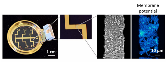
Figure 1. A) electrode dish for microbial electrophysiology developed in the PI’s group. B) Membrane potential of cells are measured by fluorescence microscopy.
Case funding
This project is suitable for CASE funding
Host
University of WarwickTheme
- Organisms and Ecosystems
Supervisors
Project investigator
Dr Munehiro Asally (Warwick) [email protected]
Co-investigators
Prof Gary Bending (Warwick) [email protected]
Dr Magdalena Karlikowska (Cytecom Ltd) [email protected]
How to apply
- Each host has a slightly different application process.
Find out how to apply for this studentship. - All applications must include the CENTA application form. Choose your application route
Methodology
Membrane potential dynamics will be measured by fluorescence time-lapse microscopy. The fluorescence intensities will be quantified for each cell using a deep-learning image segmentation tool. The change in membrane potential will be estimated in millivolt using the established equation based approaches (Ehrenberg et al., 1988). Microbial cells will be treated with chemical stresses using inhibitors, and environmental stresses such as change in temperature and water availability, and cell vitality and viability will be measured using the electrophysiological assay (Cytecom), Flow cytometry, ATP assay and colony forming unit assay. For objective 3, the growth of mycelial cells in externally applied electric fields will be imaged by microscopy using the gold-electrode dish (Figure 1). Growth of mycelia in soil and on agar will be determined following application of electric fields and growth responses imaged using a time-lapse assay.
Training and skills
Students will be awarded CENTA2 Training Credits (CTCs) for participation in CENTA2-provided and ‘free choice’ external training. One CTC equates to 1⁄2 day session and students must accrue 100 CTCs across the three years of their PhD.
- Electrical stimulation assay (microbial electrophysiology assay)
- Time-lapse fluorescence microscopy
- Macroscopic imaging
- Quantitative image analysis
- Flow Cytometry
- ATP enzymatic assays
- Plate assays
Partners and collaboration
In summary, Cytecom will make the following contributions.
- £3,500 (£1,000 per year) supplement to the Research and Training Support Grant for the duration of the studentship
- £1,000 to cover expenses associated with the 3-month industrial placement
- In-kind contributions:
- The CyteCount machine (RRP £25,000) will be provided free of charge for the duration of the project.
- 10% discount on the CyteCount consumables for the duration of the project
- 10% discount on the CytePulse consumables for the duration of the project
- Product development and data analysis support from Cytecom’s Team with a total value of £30,000
Further details
Further details on how to contact the supervisor for this project and how to apply for this project can be found here:
For any enquiries related to this project please contact Dr Munehiro Asally (Warwick) [email protected].
To apply to this project:
- You must include a CENTA studentship application form, downloadable from: CENTA Studentship Application Form 2024.
- You must include a CV with the names of at least two referees (preferably three) who can comment on your academic abilities.
- Please submit your application and complete the host institution application process via: https://warwick.ac.uk/fac/sci/lifesci/study/pgr/studentships/nerccenta/ Complete the online application form – selecting course code P-C1PB (Life Sciences PhD); from here you will be taken through to another screen where you can select your desired project. Please enter “NERC studentship” in the Finance section and add Nikki Glover, [email protected] as the scholarship contact. Please also complete the CENTA application form 2024 and submit via email to [email protected]. Please quote CENTA 2024-W14 when completing the application form.
Applications must be submitted by 23:59 GMT on Wednesday 10th January 2024.
Possible timeline
Year 1
Electrical stimulation assays with isolated strains. Detailed characterisation of membrane potential dynamics of isolated and environmental microbes.
Year 2
Detailed comparison of cell vitality and viability assays (electrophysiology, flow cytometry, ATP, plate assay)
Year 3
Characterisation of the effects of external electric field on growth polarisation
Further reading
(Gruler and Gow, 1990; Gow, 2009; Chang and Minc, 2014; Stratford et al., 2019; Benarroch and Asally, 2020; Adee, 2023)
Journal articles:
Benarroch, J. M. J. M. and Asally, M. (2020) ‘The Microbiologist’s Guide to Membrane Potential Dynamics’, Trends in Microbiology, 28(4), pp. 304–314. doi: 10.1016/j.tim.2019.12.008.
Chang, F. and Minc, N. (2014) ‘Electrochemical Control of Cell and Tissue Polarity’, Annual Review of Cell and Developmental Biology, 30(1), pp. 317–336. doi: 10.1146/annurev-cellbio-100913-013357.
Ehrenberg, B. et al. (1988) ‘Membrane potential can be determined in individual cells from the nernstian distribution of cationic dyes’, Biophysical Journal, 53(5), pp. 785–794. doi: 10.1016/S0006-3495(88)83158-8.
Gow, N. A. R. (2009) ‘Transhyphal Electrical Currents in Fungi’, Microbiology. doi: 10.1099/00221287-130-12-3313.
Gruler, H. and Gow, N. A. R. (1990) ‘Directed Growth of Fungal Hyphae in an Electric Field’, Zeitschrift für Naturforschung C, 45(3–4), pp. 306–314. doi: 10.1515/znc-1990-3-427.
Stratford, J. P. et al. (2019) ‘Electrically induced bacterial membrane-potential dynamics correspond to cellular proliferation capacity.’, PNAS, 116(19), pp. 9552–9557. doi: 10.1073/pnas.1901788116.
Book:
Adee, S. (2023) We are electric: The new science of our body’s electrome. Canongate Books.